欢迎访问南宁景明数据存储管理有限公司,我司专注生产锥齿轮、伞齿轮多年!

专业锥齿轮|伞齿轮实体厂家
专业生产各种锥齿轮、伞齿轮产品配件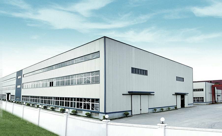
南宁景明数据存储管理有限公司
Yongjia Hongyu Gear Co., Ltd.
南宁景明数据存储管理有限公司位于浙江永嘉浦二后垟西路8号, 是一家专注生产2-15模数精密螺伞齿轮、锥齿轮多年的厂家。本公司以 “以质为尊、信誉至上”的办厂原则欢迎社会上新老客户携手合作.
质量把控

品质可靠

提供运输方案

贴心售后
宏宇齿轮 3大优势 为您保驾护航
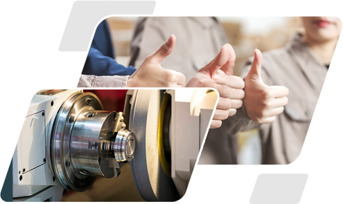
我公司拥有雄厚的技术力量、精良的设备、精湛的生产工艺
拥有可靠的生产线
Have a reliable production line and independent mold department
拥有国内外“CNC数控机床、铣齿机、研磨机、检查机、内外磨床等各式专业齿轮加工设备30多台,实力雄厚、让您合作的安心!
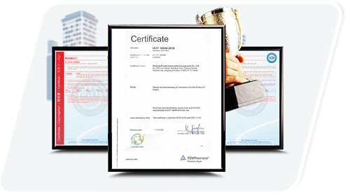
严谨的品质检验体系,设备精良
Rigorous quality inspection system, reliable materials
企业集生产,科研,销售,服务于一体,设备精良,设计工艺先进,生产及检验手段完备。公司配备精良的齿轮加工设备,本公司以 “以质为尊、信誉至上”的办厂原则欢迎社会上新老客户携手合作
提供运输方案,贴心售后
Provide efficient transportation solutions
公司有着成熟的销售方案,从咨询、定制、生产、运输、售后等一系列流程有着丰富的经验。
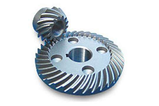
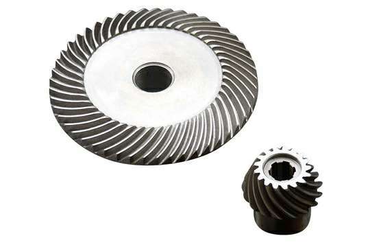
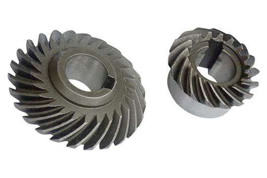
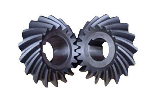
宏宇专业锥齿轮、伞齿轮生产厂家
厂家直销 / 货源充足 / 专注品质 / 诚信服务
全国销售热线
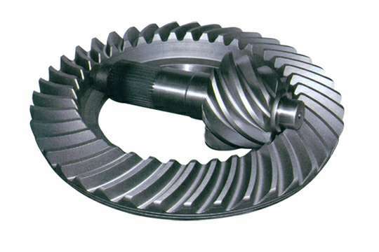
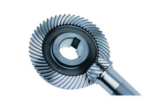
锥齿轮的优势
螺旋锥齿轮传动效率高,传动比稳定,圆弧重叠系数大,承载能力高,传动平稳,工作可靠,结构紧凑,节能省料,节省空间,耐磨损,寿命长,噪音小。在各种机械传动中,以螺旋伞齿轮的传动效率为最高,对各类传动尤其是大功率传动具有很大的经济效益;传递同等扭矩时需要的传动件传动副最省空间,比皮带、链传动所需的空间尺寸小;螺旋伞齿轮传动比永久稳定,...螺旋伞齿轮的国际标准规格要求
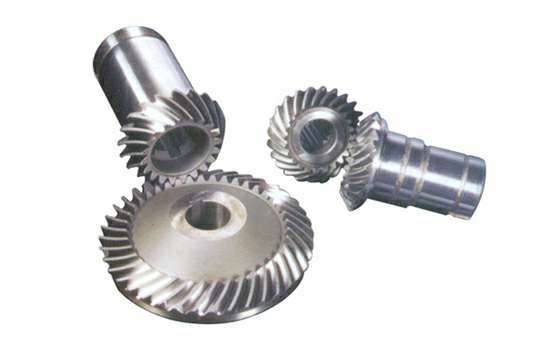
螺旋伞齿轮的国际标准规格要求:有直径10mm-1600mm、模数1-36模数、螺旋伞齿轮的精度等级有GB5级-GB8级。按照螺旋伞齿轮的设计方法、加工方法、和加工设备的不同,螺旋伞齿轮可以分为德国克林贝格螺旋伞齿轮,美国格里森螺旋伞齿轮,瑞士奥林康螺旋伞齿轮,通常被称为克林贝格螺旋伞齿轮,格里森螺旋伞齿轮和奥林康螺旋伞齿轮。这几种不同类型的螺旋伞齿轮...伞齿轮的特点和功用
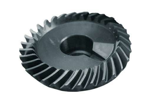
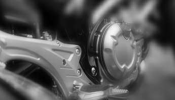
伞齿轮是齿轮中的的一种在各个工业设备中被广泛使用,下面来介绍一下它。 伞齿轮即锥形齿轮,锥齿轮。伞齿轮最常用作两垂直轴的传动,但也适应其他角度的两轴的传动。最典型的应用是用一台水平驱动装置驱动一台立式泵。在伞齿轮和正齿轮之间的两个主要区别是它们的形状和它们所在轴的关系。伞齿轮在形状上是圆锥形,而正齿轮本来是一个圆柱体。伞齿轮传递两个成角度的轴即交叉轴间的运动,而正齿轮传递两个平行轴间的运动。 圆锥齿轮的特点 锥齿轮主要用于实现两相交轴之间的运动和动力的传动,两轴交角∑称为轴角,∑的值可以根据传动的需要确定,通常多采用90度,即垂直相交。锥齿轮的轮齿排列在截圆锥体上,因此,对应于圆柱齿轮中的各有关“圆柱”的表述就变成了“圆锥”,如分度圆锥、节圆锥、基圆锥、齿顶圆锥等;这也是圆锥齿轮名称的由来。 主要特点和功用 在各种机器上所用的机械传动方法很多,最主要的有皮带传动、链条传动、摩擦轮传动、齿轮传动及绦杠螺母传动等。 其中齿轮传动,一般说来,也就是使得一个轴在旋转时能够带动另一个轴旋转;或者是把一个轴的旋转运动变为直线运动。 它的主要特点是:互相之间有齿紧凑的啮合,所传递的转矩要比皮带及链条传动大得多;它的传动效率也比其他机械传动的高;并且能在很大的传动力下保持两轴之间的速比不变。齿轮的型式甚多,大体上根据齿面形状的不同,分为圆柱齿轮、圆锥齿轮和蜗轮等型式。常用的正齿轮和斜齿轮为圆柱齿轮,它们是用来传动两个相互平行轴的旋转运动,而伞齿轮即(圆锥齿轮)是用在传动相交两轴的旋转运动的。当一对伞齿轮的轮齿啮合着传动的时候,情形和两个半截圆锥形摩擦轮的传动很相似,如图3。但是,摩擦轮的传动,如果从动轴上所带动的力量大于两轮间的摩擦力,则两轮面将会发生滑动的现象,或者甚至于使一从动轮不能被带动。如果把摩擦轮做成有齿的轮子,借齿轮上牙齿的力量去传动另一轴上的齿,就可以把这一轴的旋转力量传到另一轴上去,如图4。伞齿轮的啮合传动,就是基于这个基础上的。 伞齿轮的用途很广,尤共是遇到两轴相交、两轴的距离很近、传动的力量又很大、转数比又耍固定的时候,采用伞齿轮最为适合。